Breast Ultrasound & Automated Breast Imaging in Alberta
Breast ultrasound uses high-frequency sound waves to image internal tissues and structures of the breast. In contrast to mammography, this non-invasive procedure uses zero radiation and is safe for pregnant or breastfeeding women. Breast ultrasound is the most accessible exam of choice for any patients who have dense breast tissue, are under the age of 35, or are pregnant.
This exam is often requested as an additional test in conjunction with mammography images. There are many reasons why your doctor may send you for a breast ultrasound. Do not be alarmed if we ask to perform a breast ultrasound following or instead of a screening mammogram; often we are examining nearby lymph nodes, fluid-filled lumps (cysts), or characterizing lesions seen on the mammogram.
Purpose and Results for Breast Ultrasound
Diagnosing Breast Abnormalities
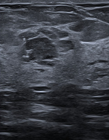
Breast ultrasound is conventionally used to investigate lumps found through self-examination, physical examination, screening mammogram, or even breast MRI. This exam helps our radiologist identify and assess the characteristics of your lesion (solid, fluid-filled, malignant, or benign) and determine the best course of action. Additionally, this exam can help explain abnormalities such as unusual nipple discharge, inflammation, breast pain, or discolouration.
Ultrasound-guided Breast Biopsy
Breast ultrasounds can be used to precisely identify a location for biopsy, so an accurate sample of the breast tissue or lump can be extracted and examined. Since an ultrasound provides images in real-time, our radiologist can rely on an ultrasound-guided breast biopsy to plan and execute the procedure in a more time efficient and less invasive way.
Mammography & Supplementary Screening for Breast Cancer Detection
Mammography is one of the best breast cancer screening methods available for early detection. Unfortunately, human physiology makes some abnormalities and lesions more difficult to detect or interpret through mammography. In particular, dense breasts with a significant amount of glandular and connective tissue can disguise potential concerns. For this reason, we may perform supplementary imaging using breast ultrasound or MRI.
Although MRI provides a higher level of detail than conventional ultrasound, it might not be available or needed in all cases. Our radiologist will work with your doctor to decide the best course of action.
ABUS: Automated Breast Ultrasound & Breast Imaging in Alberta
ABUS is specifically designed for detecting cancer in dense breast tissue by providing a comprehensive view of the breast. With mammography alone, dense breast tissue can make it more difficult to identify the appearance of tumours, which is why your doctor may recommend ABUS.
In average-risk women with dense breasts, adding ABUS to routine screening mammography increases the detection of breast cancer by at least 36% (likely more). These cancers tend to be small, invasive, and node-negative meaning they haven’t spread to the lymph nodes. Because ABUS is automated, it decreases your examination time.
Currently offered at our Hermitage, Meadowlark, Lendrum, and Millwoods locations, ABUS is a painless exam that will take approximately 20-30 minutes to complete.
For more information about ABUS, click here.
Diagnostic Breast and Axilla Ultrasound Exam
Diagnostic breast and axilla ultrasounds produce images that show abnormalities within the breast and armpit, scientifically known as the axilla.
More specifically, this form of imaging examines the concentration of lymph nodes in your armpit, looking for irregularities and helping identify abnormal axillary sentinel lymph nodes.
Lymph node status is critical when evaluating medical or surgical therapies. Since your axilla has the largest concentration of lymph nodes in the body, symptoms are more likely to appear there first.
FAQ
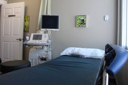
- You will be asked to change into a gown, removing your clothing from the waist up.
- You will need to expose your breast for the exam so the sonographer can scan it in its entirety, along with surrounding tissue and the axilla (armpit).
- A warm, hypoallergenic ultrasound gel will be applied to the area being scanned. The sonographer will glide a probe over your breast and the surrounding area to provide the radiologist with a complete set of images.
- Insight Medical Imaging only uses female sonographers for breast and axilla ultrasounds, so our patients are more comfortable when their breast is exposed.
- The sonographer will apply moderate pressure with the probe, which may be momentarily uncomfortable, but it should not cause any pain.
- After the sonographer has captured the images, you are free to leave.
- The results will be analyzed by one of our radiologists and a detailed report will be sent to your doctor. We try our best to send the results as soon as possible, usually within one business day.
This procedure does not use radiation; therefore, it poses no direct threat to pregnant women. While different health conditions may predispose you to certain risks, the primary risk in breast ultrasound is that imaging may not show extremely tiny lesions and will not identify tiny calcifications often associated with node negative precancers.
Obese patients, or those with abnormally large breasts are at higher risk for misdiagnosis, as there is a negative correlation between body mass or size of breasts and ultrasound accuracy. Therefore, it is highly likely for your doctor to refer you for screening mammography before an ultrasound, for better lesion localization accuracy.
Diagnostic breast and axilla ultrasound exams typically take 30 minutes per side. Sometimes our team will scan both sides, and in this case, the exam may take longer than 30 minutes. ABUS Breast screening usually takes about 20-30 minutes in total to complete. If you have more questions about your breast ultrasound exam, please call 1-866-771-9446 to speak with one of our health technologists.
ABUS is used for detecting and better understanding breast lesions that may be present in a patient, and it is usually used in conjunction with other breast screening and imaging methods, like mammograms. Automated Breast Ultrasound is especially beneficial and often recommended for patients with higher breast density since it increases the likelihood of tumor and cancer detection by at least 36%. If you have a dense breast area and would like more information, please contact our Hermitage and Spruce Grove locations to find out how ABUS can increase the accuracy of your breast cancer assessment.
Automated Breast Ultrasound, or ABUS, is a new, non-invasive breast imaging system. This imaging technology, much like a normal ultrasound, uses sound waves to create a 3D image of the entire breast. ABUS does not use any radiation, so the test is safe for pregnant women or those with conditions that may exclude them from tests involving radiation.
ABUS differs from ultrasound in a few ways. First, ABUS uses a larger, automated probe to generate images of the breast tissue, meaning no operator is required to hold the ultrasound probe. Second, ABUS automatically captures hundreds of thin images of the breast, rendering them into a 3D image, instead of ultrasound’s 2D image, showing the breast tissue in greater detail.
No, ABUS is not a replacement for a mammogram. ABUS, like a normal ultrasound, is used in conjunction with a mammogram to better detect early signs of cancer. For women with dense breast tissue, it may be more difficult to detect cancer, so a physician might also recommend an ABUS test.
You will need to remove your clothing from the waist up and wear a robe for comfort. You will be asked to lie on your back and warm gel will be applied to your chest/breast area. A technologist will then use a transducer to scan the chest and underarm area. You might feel some pressure during the exam, but it should not be painful and is completely non-invasive. Your technologist might focus on particular areas to get comprehensive images. Once the practitioner captures enough images, you can wipe off the gel and dress.
Cost
If you have an Alberta Health Care card or valid healthcare card from out of province, there is no cost for a breast and axilla ultrasound (except Quebec).
Exam Preparation
Being prepared for your breast ultrasound helps us take the best possible images for diagnosis. Please visit our exam prep page for more instructions specific to breast and axilla ultrasound.