Vascular Ultrasound
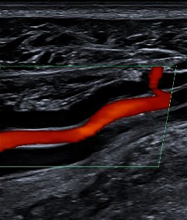
Vascular ultrasound is a non-invasive form of diagnostic imaging that uses high-frequency sound waves to assess the circulation of blood throughout the human body. There are numerous types of vascular ultrasound exams available, but the most common are carotid, peripheral venous (DVT), and peripheral arterial (ABI) for arms and legs.
Reasons for a Vascular Ultrasound
Your doctor may refer you for a vascular ultrasound to assess your circulatory system. More specifically, this exam evaluates the flow of blood through your veins and arteries to different parts of your body, looking for blockages, clots, or abnormalities.
Your doctor will also rely on a vascular ultrasound to:
- Get information about blood supply to organs like the brain
- Locate stenosis, plaque buildup, or narrowing of blood vessels
- Evaluate if you are eligible for angioplasty or a stent
- Assess varicose veins
- Identify localized weaknesses in blood vessel walls
Orientation
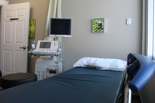
During a vascular ultrasound, you will be on a bed, on your back. Our sonographer may ask you to change position during your scan, depending on the area of interest (legs, arms, neck, etc.)
Cost
If you have an Alberta Health Care card or valid health care card from out of province, there is no cost for a vascular ultrasound (except in Quebec).
Duration
A vascular ultrasound scan lasts approximately 30 to 60 minutes, depending on the scan location.
- A carotid ultrasound lasts approximately 45 minutes.
- A peripheral venous ultrasound of the arm lasts about 45 minutes.
- A DVT of the leg lasts about 30 minutes.
- A peripheral arterial ultrasound of the arm lasts about 60 minutes.
- An ABI of the leg lasts about 30 minutes.
Common Vascular Ultrasound Assessments:
Carotid Ultrasound
Carotid ultrasound exams use sound waves to assess blood flow through the two carotid arteries in the neck. These arteries are crucial, as they deliver blood from the heart to the brain.
Why Would I Need a Carotid Ultrasound?
Carotid ultrasounds inform your doctor about stenosis and the health of your arteries. Testing for narrowed arteries helps your doctor assess your risk of stroke. An early diagnosis can give you enough time to implement a medical or surgical treatment plan to reduce or remove blockages in your arteries.
Candidates for Carotid Ultrasound
Some reasons your doctor may refer you for a carotid ultrasound include:
- High blood pressure / cholesterol
- Diabetes
- History of heart disease
- Recent transient ischemic attack (TIA)
- Coronary artery disease
- Recent mini-stroke
What Happens During My Carotid Ultrasound?
- You will lie on a bed, and the clothing around your neck will be removed.
- A warm, hypoallergenic ultrasound gel will be applied to your neck.
- The sonographer will move the transducer (probe) along both sides of your neck, from your jaw to your collarbone.
- The pressure should not cause pain. Please inform your sonographer of any discomfort.
- The ultrasound will capture images of blood vessel structure and blood flow to the brain.
- After the sonographer has captured the images, you are free to leave.
- A radiologist will review the images and send a detailed report to your doctor, usually within one business day.
Peripheral Venous (DVT) Ultrasound
DVT ultrasounds use high-frequency sound waves to produce images of your body’s extremities – arms and legs.
Why Would I Need a DVT Ultrasound?
DVT ultrasounds are most commonly ordered to search for clots in the veins of your leg. Your doctor may refer to this exam as a deep-vein thrombosis or DVT ultrasound.
Candidates for DVT Ultrasound
Your doctor may refer you for a DVT ultrasound to investigate:
- Swelling or redness of a limb
- Chronic venous insufficiency
- Vascular malformations or varicose veins
- Deep-vein thrombosis
- Superficial thrombophlebitis
- Blood vessel grafts used for dialysis
What Happens During My DVT Ultrasound?
- Once you arrive, we may ask you to change into a gown or to remove your pants or top, depending on whether we are examining your arm or leg.
- The sonographer will apply a warm, hypoallergenic ultrasound gel and move the transducer (probe) along your leg or arm.
- If the sonographer is scanning your leg, they will move the probe from your groin to your knee.
- The sonographer will scan each segment of the vein down your leg, looking for blood clots and buildups.
- If the sonographer is scanning your arm, they will move the probe from your neck down to your elbow (and occasionally your wrist).
- Closing the veins momentarily from the pressure should not cause pain but will likely be uncomfortable. This discomfort is normal, but please inform your sonographer of any pain.
- The sonographer may reposition you during the scan to capture better images.
- After the sonographer has captured the images, you are free to leave.
- A radiologist will review the images and send a detailed report to your doctor, usually within one business day.
Peripheral Arterial (ABI) Ultrasound
ABI ultrasounds use high-frequency sound waves to assess the blood flow between your arms, groin, legs, and ankles. During an ABI ultrasound, our sonographer will also measure the difference in blood pressure between your arm and ankle, which is known as an ankle-brachial index or ABI ultrasound.
Why Would I Need an ABI Ultrasound?
Your doctor will refer you for an ABI ultrasound to learn more about the circulation of blood through the arteries in your arms, legs, and ankles. Unlike a DVT ultrasound, this exam does not look for blood clots in veins, but rather blood vessel narrowing.
Candidates for ABI Ultrasound
Your doctor may refer you for an ABI ultrasound to investigate:
- Resting leg pain or muscle cramps
- Difficulty walking long distances
- Ulcers in the foot, ankle, or heel
What Happens During My ABI Ultrasound?
- You may be asked to change into a gown for access to your legs and arms.
- The sonographer will position you on your back and attach blood pressure cuffs to both arms and ankles.
- The blood pressure cuffs will inflate so the sonographer can take your blood pressure readings.
- You will feel some pressure for a short time while the cuffs inflate.
- The sonographer will apply a warm, hypoallergenic ultrasound gel to both arms/legs (the area of interest).
- During the exam, your sonographer will listen to your blood flowing through each artery, which will sound like a heartbeat.
- After the sonographer has captured the images, you are free to leave.
- A radiologist will review the images and send a detailed report to your doctor, usually within one business day.
Exam Preparation
Being prepared for your vascular ultrasound helps us take the best possible images for diagnosis. Visit our exam prep page for more instructions specific to vascular ultrasound.