
Why is Compression Important for a Mammogram?
June 16, 2019

Most women know how important it is to schedule a screening mammogram every two years once they turn 50. The Canadian Cancer Society recommends you talk to your doctor about screening options as soon as you turn 40.[2] At Insight Medical Imaging, we recommend you strongly consider annual screening mammograms once you turn 40, especially if you have any of the risk factors associated with breast cancer or a family history of breast cancer.
In 2017, Statistics Canada noted that only 78.5% of women received a screening mammogram in the past two years. The main reasons for not getting a mammogram include: “did not think it was necessary,” “did not get around to it,” or were simply afraid (of the pain, embarrassment, or fear for positive test results).[3]
Screening mammography is such a vital part in the fight against breast cancer. Although mammograms may cause momentary discomfort for women with sensitive breasts, the positives far outweigh the negatives. While the idea of breast compression sounds scary, it’s crucial for image quality and diagnosis.
Breast Compression and Abnormality Detection
To detect breast abnormalities, compression is needed to reduce the thickness of the breast. While it is widely accepted that breast compression improves image quality, there is no industry standard indicating how much compression should be applied during the mammogram.[4]
If a breast is not well compressed, overlapping tissue can appear similar to an abnormality, leading to a false positive, or a mass could be missed altogether. A compressed breast also requires less radiation exposure to generate high-quality images and reduces motion-produced image discrepancies generated from patient movements.
Mammography at Insight Medical Imaging
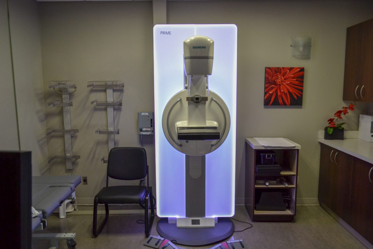
When you visit Insight Medical Imaging for your screening mammogram, our technologists will help position your breast on one of our breast tomosynthesis units (3-D mammography). The breast tissue will be gently compressed for a short duration, approximately 5 to 40 seconds per image.
The variance in compression is based on the machine’s tomosynthesis time. At least two images per breast will be taken for a screening mammogram. If you return for a diagnostic mammogram, we may take additional photos of each breast to help our radiologists with diagnosis.
Sometimes our team will also recommend performing a breast ultrasound after your mammogram to provide your doctor with even more information about the health of your breasts.
While 40 seconds for a single compression may seem long, it is important to remain as still as possible. Our special breast tomosynthesis mammography machines examine breast tissue layer by layer in a three-dimensional (3-D) plane. By imaging your breast in thin layers, our radiologists can obtain a higher-quality, clearer, complete picture of your breasts.
Additionally, breast tomosynthesis technology has been shown to increase breast cancer detection in early stages and reduce the need for additional imaging.[8] This means faster appointments for you and a smaller chance that you will require additional imaging.
Hologic Breast Tomosynthesis Systems
Insight Medical Imaging has also invested in Hologic Breast Tomosynthesis SmartCurve Sytems that use a curved compression plate that mirrors the shape of a woman’s breast to reduce pinching. The system applies uniform compression over the entire breast for added comfort.
The SmartCurve System has been shown to improve comfort in 93% of patients who reported moderate to severe discomfort with standard compression technology,[8] while still providing our radiologist and your doctor with high-quality images. This technology allows us to increase comfort while we image your breasts, while reducing the chance that you might need follow-up visits.
We understand going for a mammogram can be stressful. That’s why our team does everything in our power to make your visit as pleasant as possible. We recommend visiting our dedicated breast imaging facility, Lendrum Women’s Imaging, for your next breast imaging exam. Our staff is highly experienced in diagnostic women’s imaging and look forward to helping you.
References
- Agasthya, G., D’Orsi, E., Kim, J., Handa, P., Ho, C., D’Orsi, C., Sechopoulos, I. (2017). Can Breast Compression Be Reduced in Digital Mammography and Breast Tomosynthesis? Retrieved from https://www.ajronline.org/doi/10.2214/AJR.16.17615.
- Canadian Cancer Society. (2017). Mammography. Retrieved from http://www.cancer.ca/en/cancer-information/diagnosis-and-treatment/tests-and-procedures/mammography/?region=on.
- Canadian Cancer Society. (2017). What is Breast Cancer? Retrieved from http://www.cancer.ca/en/cancer-information/cancer-type/breast/breast-cancer/?region=ab.
- Holland, K., Sechopoulos, I., Mann, G., Van Gils, C., Karassemeijer, N. (2017). Influence of Breast Compression Pressure on The Performance of Population-Based Mammography Screening. Retrieved from https://www.ncbi.nlm.nih.gov/pmc/articles/PMC5706300/.
- Kennedy, S. (2018). Why is compression important during a mammogram? Retrieved from https://www.wakerad.com/expert-feature/why-compression-is-important-mammogram/.
- Poulos, A., McLean, D., Rickard, M., Heard, R. (2003). Breast Compression in Mammography: How Much is Enough? Retrieved from https://www.ncbi.nlm.nih.gov/pubmed/12780439.
- Poulos, A., McLean, D. (2004). The Application of Breast Compression in Mammography: A New Perspective. Retrieved from https://www.radiographyonline.com/article/S1078-8174(04)00043-4/abstract.
- Smith, A. (2017). Improving Patient Comfort in Mammography. Hologic, Rev 003.
- Statistics Canada. (2018). Health Fact Sheets – Cancer Screening, 2017. Retrieved from https://www150.statcan.gc.ca/n1/pub/82-625-x/2018001/article/54977-eng.htm