
MRI: What It Tells You About Your Brain
July 3, 2023
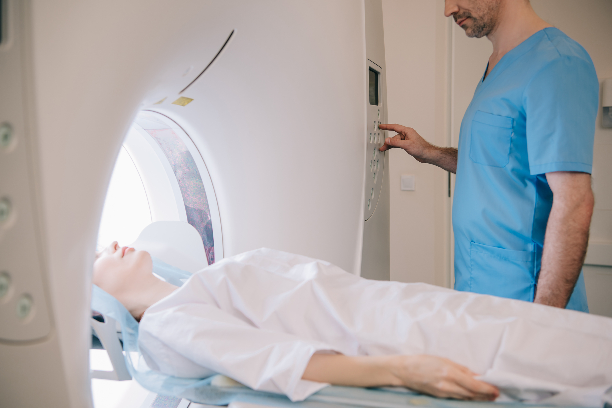
MRI stands for Magnetic Resonance Imaging. It is a non-invasive diagnostic tool that uses a magnetic field, radio waves, and a computer to produce detailed images of the organs and tissues within the body. These images can be used to diagnose and monitor a wide range of medical conditions, including tumours, blood vessel diseases, infections, and injuries. MRI is particularly useful for imaging the brain and spinal cord, as well as the joints, and other internal organs. Because it does not use ionizing radiation, it is considered a safer alternative to X-rays and CT scans.
Why would a neurologist order an MRI?
A neurologist might order an MRI of the brain to diagnose or evaluate a wide range of neurological conditions such as traumatic brain injury, stroke, tumours, multiple sclerosis, infections, epilepsy, dementia and other degenerative conditions. An MRI can help detect bleeding or swelling in the brain, identify the area affected by the stroke, detect the presence and location of brain tumours, evaluate the severity of the disease and monitor its progression over time, detect brain abscesses and other infections, identify the location of the seizure focus in the brain and aid in the diagnosis of the cause of seizures, as well as identify brain changes associated with dementia and other degenerative conditions. It may also be used to evaluate symptoms such as headaches, changes in vision, and changes in cognitive function.
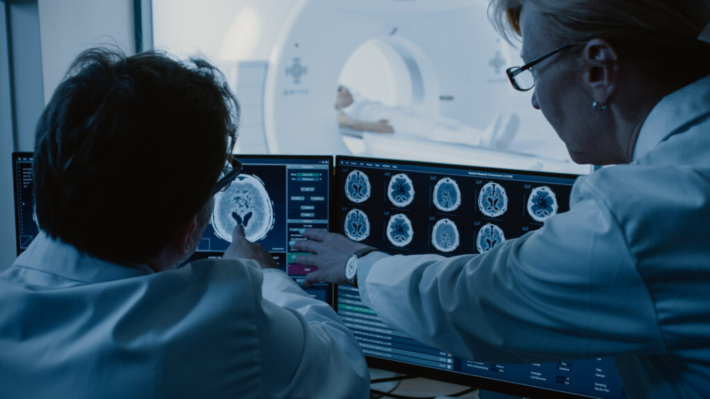
An MRI can provide detailed images of the brain and can show a variety of information about the brain’s structure and function. An MRI can show:
- The size, shape, and position of brain structures, such as the brainstem, cerebellum, and cerebral hemispheres.
- The presence of any abnormal growths or tumours, such as brain tumours or cysts.
- Damage or abnormalities in the brain tissue, such as from a stroke or traumatic brain injury.
- The location and size of any fluid-filled spaces, such as ventricles or cysts.
- The presence of any brain infections, such as encephalitis or meningitis.
- The presence of any degenerative conditions, such as Alzheimer’s disease or multiple sclerosis.
- The size and shape of the brain ventricles, which can indicate the presence of hydrocephalus.
- The presence of any structural defects or malformations, such as a congenital brain malformation.
It’s important to note that an MRI can help identify potential issues, but it’s not conclusive. A neurologist or radiologist will need to interpret the results and other medical information to make a diagnosis.
How to prepare for an MRI
To prepare for an MRI scan, it is important to follow any instructions provided by the facility where the scan will be performed. Generally, you should wear comfortable, loose-fitting clothing and remove any metal objects, such as jewelry or hairpins, that may interfere with the scan. You may also be asked to remove any clothing with metal zippers or snaps. You may also be asked to refrain from eating or drinking for a certain period of time before the scan. If you have any concerns about the scan or are pregnant, it is important to inform the facility before the scan.