Mammography
Mammography uses a low-dose X-ray to either screen for or diagnose breast disease. The term mammogram refers to the image captured during mammography. These images help our team look for malignant and benign abnormalities in the breast before physical symptoms are present. In the fight against breast disease, early detection is crucial.
Insight Medical Imaging recommends breast cancer screening guidelines from the Canadian Association of Radiologists (CAR), the American College of Radiology (ACR) & the Society of Breast Imaging (SBI) as well as the National Comprehensive Cancer Network (NCCN). These guidelines educate practitioners, radiologists, and technologists so there is a consistent understanding of what is considered optimal patient care. These guidelines are not binding for practitioners, but rather evidence-informed principles from leading breast cancer experts to help detect cancer early when it is small, easier to treat, and less likely to be fatal.
Recommendations for Asymptomatic, Average Risk Women are:
< 40 years old: Screening is not routinely recommended because the incidence of breast cancer is low in this age group
40-49 years old: Annual Screening Mammography is recommended. Women in this age group tend to have higher breast density (which can obscure small early cancers) and cancers tend to grow more rapidly.
50-74 years old: Both the ACR/SBI and the NCCN recommend Annual Screening Mammography whereas the CAR recommends every 1-2 years (biennial). Insight recommends annual screening mammography to be strongly considered but these women should never go longer than 2 years between screening. Annual mammography decreases a woman's chance of dying from breast cancer compared to biennial screening, particularly for those with dense breasts.
>74 years old: Both the ACR/SBI and the NCCN recommend continuing with Annual Screening Mammography whereas the CAR recommends continuing with screening every 1-2 years (biennial). However, if a woman has severe comorbidities (medical problems) expected to limit her life expectancy, then screening mammography is not recommended.
Alberta Mammography
With Testing Locations in Edmonton, St. Albert, Fort McMurray, Leduc, Sherwood Park & Spruce Grove
1) Screening Mammogram
A screening mammogram uses low-dose X-rays to detect abnormalities within men’s and women's breasts that are too small to be felt as a lump by a patient or doctor. The goal is to detect breast cancer in its earliest stage without involvement of the lymph nodes, significantly increasing chances of successful treatment.
Insight Medical Imaging follows the Alberta Breast Cancer Screening Program (ABCSP), which is coordinated by Alberta Health Services and the Alberta Society of Radiologists (ASR). The program’s purpose is to educate the public about imaging options and why regular screening mammograms are so important in the fight against breast cancer.
As part of the ABCSP regulations, patients can self-refer for a screening mammogram once a year, provided their first screening exam had a doctor’s referral and they have no symptoms at the time of the exam.
Typically, the governing body for diagnostic imaging states that all patients must have a doctor’s referral for imaging exams. However, screening mammograms are an exception to this rule. As the ABCSP states, “just because no one in your family has had breast disease doesn’t mean you’re not at risk.”
Learn more about breast disease risk factors you can and cannot control and how to assess your risk, and get helpful information about breast cancer screening from Screening for Life.
2) Diagnostic Mammogram
A diagnostic mammogram is similar to screening as it also uses low-dose X-rays to investigate abnormal clinic findings or unusual symptoms discovered by your doctor. These symptoms range from tenderness, skin changes, a lump, nipple discharge, etc. Diagnostic mammograms can also follow screening mammograms, be prescribed as an interval follow-up, and confirm whether a finding is benign or if additional testing is required.
Digital Breast Tomosynthesis
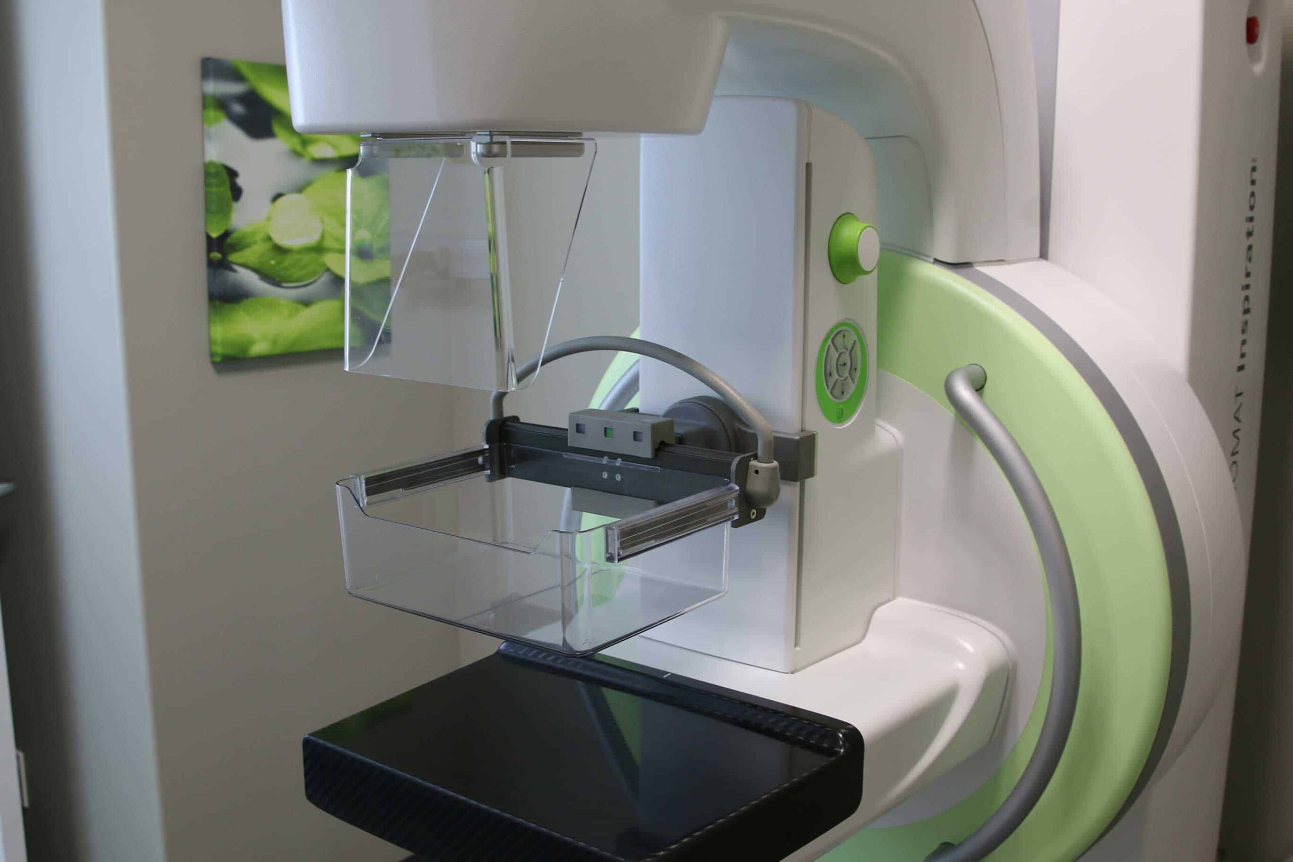
Insight Medical Imaging uses digital breast tomosynthesis (DBT) when imaging your breasts during a mammogram. Breast tomosynthesis is an advanced form of imaging that examines the breast tissue layer by layer in a 3-D plane, as opposed to a standard mammography exam which examines in 2-D.
During the procedure, an X-ray arm moves over the compressed breast, capturing images of the tissue from various angles. These images are then processed or “synthesized” into a 3-D picture by a computer. The result is a very detailed set of 3-D images, which our radiologist can examine from any angle to distinguish normal overlapping tissue that might otherwise be misdiagnosed as an abnormality.
This technology provides our radiologists with higher-quality images that make it easier to detect small abnormalities. Since breast tomosynthesis provides such detailed images, it is less likely that additional imaging will be required. Therefore, our patients are not exposed to unnecessary radiation or stress.
What Happens During my Mammogram?
- Upon checking in, you will be asked to complete an ASR patient information form that contains questions about your health and history.
- Next, you will be asked to change into a gown, removing your clothing from the waist up.
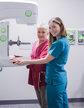
- A technologist will help position you in front of the machine, with your breast between two X-ray plates.
- To ensure our patients are as comfortable as possible during their mammogram, only female technologists scan when a breast is exposed.
- The surfaces will slowly move together, compressing the breast tissue for a short duration.
- Please be assured that only the necessary amount of compression is applied for optimal image clarity and sharpness. Our technologists are aware that compression is uncomfortable and will work with you to provide the best examination possible.
- We will take at least two images of each breast (top to bottom and a side view).
- We may need to take additional images or perform an ultrasound. Do not be alarmed. Often, our radiologists will request additional imaging to ensure a thorough examination.
- After the sonographer has captured the images, you are free to leave.
- The results will be analyzed by one of our radiologists and a detailed report will be sent to your doctor. We try our best to send the results as soon as possible, usually within one business day.
ABUS Breast Imaging
ABUS is specifically designed for detecting cancer in dense breast tissue by providing a comprehensive view of the breast. With mammography alone, dense breast tissue can make it more difficult to identify the appearance of tumors, which is why your doctor may recommend ABUS.
In average-risk women with dense breasts, adding ABUS to routine screening mammography increases the detection of breast cancer by at least 36% (likely more). These cancers tend to be small, invasive, and node negative.
Currently offered at our Hermitage, Meadowlark, The Grange, Lendrum Women's Imaging, Millwoods, St. Albert, and Spruce Grove locations, ABUS is a quick, non-invasive exam-taking about 20-30 minutes to complete.
For more information about ABUS, click here.
Cost
Screening Mammogram
Alberta Health covers one screening mammogram per calendar year (January -December). For example: if you have a mammogram in January, 2019, you will have to wait until at least January, 2020, before your next screening mammogram.
Diagnostic Mammogram
Your doctor has likely referred you for this exam. If you have an Alberta Health Care card or valid healthcare card from out of province, there is no cost for a diagnostic mammogram (except Quebec).
ABUS Breast Imaging
Your doctor or one of our radiologists have likely referred you for this exam. If you have an Alberta Health Care card or valid healthcare card from out of province, there is no cost for a diagnostic mammogram (except Quebec).
Duration
Both screening and diagnostic mammograms take approximately 20 minutes to complete.
Exam Preparation
Being prepared for your mammogram helps us take the best possible images for diagnosis. Please visit our exam prep page for more instructions specific to mammography.


