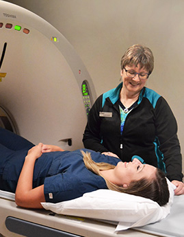
Extremity CT
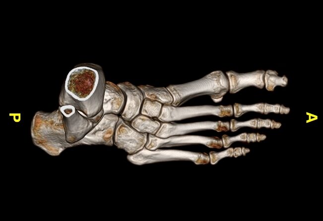
An extremity CT scan uses x-rays to create 2D and 3D images of the the extremities such as the hand and joints such as the knee. CT imaging is similar to a conventional x-ray, but the images are more detailed and can be combined by a computer to generate 3-D representations that allow our radiologist to view the area of interest from multiple angles.
Reasons for an Extremity CT Scan
Your doctor might order this imaging test to examine different types of tissue or bone structure in a perimeter body part such as your hand. This scan will provide detailed information about the bone and soft tissue in the area of interest and help screen for abnormalities.
This exam can help your doctor:
- Evaluate pain, swelling, or trauma
- Identify and localize a known mass
- Examine complex fractures
- Diagnose arthritis
- Scan for a collection of pus (abscess)
- Monitor scar tissue and healing after surgery
Lower Extremities vs. Upper Extremities
There are two main groups of extremities, upper and lower, classified by their location in the body.
Lower extremities include:
- Femur
- Knee
- Tibia and fibula
- Foot and ankle
Upper extremities include:
- Humerus
- Elbow
- Forearm
- Wrist and hand
Duration
An extremity CT scan takes less than a minute to perform. Your entire visit should take less than 30 minutes.
Exam Preparation
Being prepared for your CT scan helps us take the best possible images for diagnosis. Please visit our exam prep page for more instructions specific to extremity ct preparation.