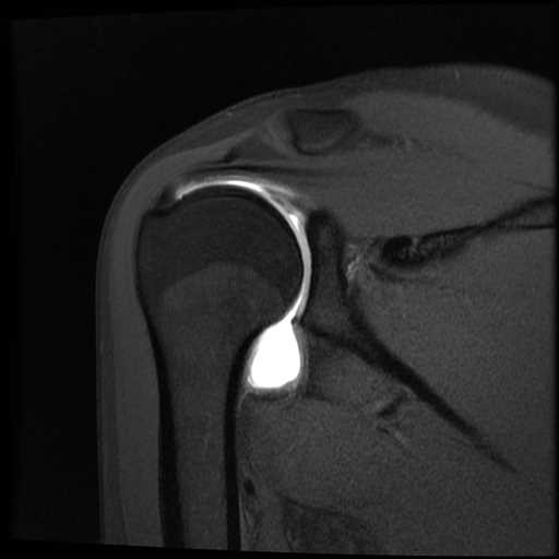
MR Arthrogram Imaging
An MR arthrogram is a study used to better visualize the structures within the joint. An MRI is performed following the fluoroscopy-guided injection of contrast directly into the joint. More commonly, it is also possible to use MRI or CT imaging to capture the tissue structures in greater detail. The images from this exam help doctors make informed decisions regarding treatment and monitor recovery and healing. Arthrograms are typically ordered to examine soft tissue inside the joint, particularly if a patient has dislocated that joint multiple times. This exam is a private service and is not reimbursed by Alberta Health.
Orientation
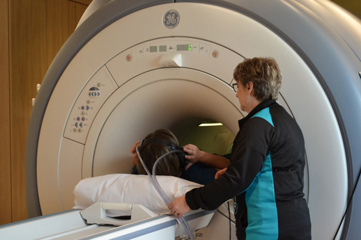
During MR arthrogram imaging, you will be moved into the MRI machine head-first. If you are claustrophobic, please consult with your doctor to see if they recommend that you take anti-anxiety medication before your exam.
Cost
Alberta Health Services does not cover private MRI services outside of the hospital. The cost of a MR arthrogram ranges from $800 to $950, depending on the need for contrast.
The need for contrast is determined by the radiologist at the time of imaging if it is determined that it is required for a proper diagnosis. The need for contrast is also impacted by specific patient history.
Duration
Part 1: Injection – 10 minutes
Part 2: MR scan – typically 30-60 minutes
The technologist, who can hear and see you during the exam, will provide you with an emergency button so you can communicate with them throughout the procedure.
Additional Procedure Details and Expectations
Part 1: Injection
- Upon being checked in, a technologist will go over a safety questionnaire with you to determine whether you are a safe candidate for the injection and MRI imaging.
- Once you arrive, you will be asked to change into a gown, unless the joint is easily accessible, such as in your ankles and hands.
- Before the start of your exam, you will fill out a short questionnaire and provide written consent for your procedure. Direct any questions you have to our technologists.
- You will be asked to lie on a table and the area to be injected will be exposed and cleaned with an aseptic
- Your doctor will use either fluoroscopy (a type of X-ray) or ultrasound to help view the area.
- The skin may be numbed with a local anesthetic and contrast may be injected into the joint to confirm needle placement. If you are allergic to contrast, your doctor will discuss your options before the procedure.
- The injection procedure takes approximately 10 minutes.
- You will be awake during the procedure and may ask questions at any point.
- During the injection, you may experience mild to moderate discomfort in the area of the injection.
- Once the injection is complete, you will be taken to the MRI department for the scan.
Part 2: MR Imaging
- We will provide you with ear protection to soften the noise of the MRI machine as well as any special equipment needed to help us obtain the best possible images.
- You will then lie on the scanning table and your head will be centred into the magnetic tunnel.
- Your exam may require contrast to provide us with optimal images. Before the exam, please let the technologist know if you’ve experienced allergies to contrast in the past. If contrast is needed, a technologist will insert an IV line into a vein in your hand or arm and inject the contrast agent. You may feel a cooling sensation.
- This exam takes approximately 30 to 60 minutes. During this time the technologist can hear and see you. All MRI exams require you to be very still in order to take good images.
- Some patients may find the MRI machine makes them feel claustrophobic, especially during a brain MRI. If you think you might have a hard time with the exam, please talk to your doctor. They may prescribe a mild sedative to help you relax during the scan.
- The results will be analyzed by our on-site radiologist and a detailed report sent to your doctor as soon as possible.
Exam Preparation
Please visit our exam prep page for more instructions specific to MR arthrography preparation.