Musculoskeletal - MSK Ultrasound
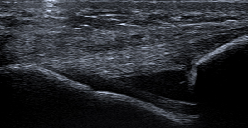
Musculoskeletal ultrasound, or MSK, is a special type of ultrasound imaging that uses high-frequency sound waves to assess joints and supporting tissue such as tendons, ligaments, muscles, and cartilage. This exam is often used to evaluate the space between your joints and interacting tissue when looking for muscle/tendon tears or for fluid buildup and inflammation that may harm mobility.
Reasons for a MSK Ultrasound
Your doctor may refer you for an MSK ultrasound to help diagnose sprains, strains, tears, or pain in your joints or soft tissue. This non-invasive form of diagnostic imaging can help determine the severity of an injury and if surgery is required, or assess how an injury is healing.
Additionally, MSK scanning is commonly used to evaluate sports-related injuries, such as tears in the rotator cuff or Achilles tendon. These tears are often hard to diagnose since the pain or symptoms are usually only present when making specific motions.
Traditional imaging like X-ray or MRI requires the patient to remain very still, making it challenging to capture intricate structures in real time without sacrificing image quality. MSK allows our radiologists to view intricate structures, such as joints, from multiple angles at the exact moment when there is pain or discomfort.
Who Is Eligible for an MSK Ultrasound?
Everyone is eligible for a musculoskeletal ultrasound if they have a physician’s referral. These exams are popular for athletes because many of their injuries are related to physical exhaustion and exercise. However, it is not uncommon to perform an MSK scan on an elderly patient to assess early arthritis development or muscular calcifications.
Common Areas of Interest
- Shoulder
- Elbow
- Wrist
- Hip
- Knee
- Ankle
- Extremities (hand, finger, foot, toe)
MSK ultrasound can also be used to investigate plantar fascia, carpal tunnel, ganglion bumps, unusual lumps on muscles or tendons, and more.
MSK Ultrasound vs. Magnetic Resonance Imaging (MRI)
Musculoskeletal ultrasound has comparable diagnostic capability to MRI when assessing traumatic, inflammatory, or degenerative soft-tissue conditions and the health of your body’s joints. While state-of-the-art 1.5 Tesla and 3 Tesla magnetic resonance units provide superior image detail, MSK ultrasound has helpful diagnostic capabilities and offers these unique benefits:
- MSK provides real-time dynamic imaging, which helps demonstrate muscle pathology. Conversely, MRI exams require patients to remain completely still for the entire exam.
- Alberta Health Services covers MSK ultrasound, and exam availability is usually better. While private MRI exams are available, the cost of the procedure is a significant deterrent for most people.
- MSK ultrasound can be safely performed on all patients whereas MRI should not be performed on patients who have pacemakers, embedded metal fragments, or surgical implants.
- MSK ultrasound is portable and significantly more comfortable than a potentially claustrophobic MRI tunnel.
While MSK ultrasound provides some unique benefits over MRI, both exams are often ordered to provide a complementary analysis. Your doctor may start with an MSK ultrasound, as typical wait times are three to four weeks.
While this may seem like a long time, public MRI exam wait times for soft tissue can extend past one year. If you do not wish to wait for a public MRI, you can book a private scan with us within a few days for a fee.
It is logical to order an MSK ultrasound to evaluate your tissue, as it can provide actionable diagnostic results months before a public MRI or save you from investing in a private scan that may be unnecessary.
What Happens During My MSK Ultrasound?
- You may be asked to change into a gown, depending on which body part we are examining. Alternatively, the sonographer may arrange your clothing to expose the area of interest so you do not have to change.
- The sonographer will apply a warm, hypoallergenic ultrasound gel and move the transducer around the area of interest with moderate pressure. This pressure should not cause pain. Please inform your technologist of any discomfort.
- The sonographer may ask you to change position so they can obtain the best possible images.
- After the sonographer has captured enough images, you are free to leave.
- A radiologist will review the images and send a detailed report to your doctor, usually within one business day.
Important Notes
It is not uncommon to X-ray an area of interest before a musculoskeletal ultrasound. Combining both sets of images (bones and soft tissue) allows our radiologists to compare results and aids in diagnosis.
Orientation
During a musculoskeletal ultrasound, your position will be based on which body part is being scanned.
If you are having an upper body part scanned, you will likely be sitting upright on a bed. If you are having a lower body part scanned, you will likely be lying on your back or side, whichever gives the sonographer better access to the area of interest.
Occasionally, you will be asked to stand for imaging. Sometimes patients are asked to flex muscles. Flexing can be difficult for some patients, which is why we position them in a weight-bearing stance, such as standing.
Cost
If you have an Alberta Health Care card or valid health care card from out of province, there is no cost for a musculoskeletal ultrasound (except in Quebec).
Duration
A musculoskeletal ultrasound lasts approximately 30 minutes for each body part being scanned.
Exam Preparation
Being prepared for your MSK ultrasound helps us take the best possible images for diagnosis. Visit our exam prep page for more instructions specific to MSK ultrasound.


