Echocardiogram in Alberta With Testing Locations in Fort McMurray, Sherwood Park & Spruce Grove
An echocardiogram, or “echo,” is a specialized ultrasound of the heart using 2-D and Doppler ultrasound. This non-invasive procedure provides information about the size and shape of the heart, its pumping capacity, and blood flow through the heart chambers. Echocardiography can also locate and evaluate heart damage, which your doctor can use to identify heart disease.
Reasons for an Echocardiogram
Your doctor may refer you for an echocardiogram to evaluate the health of your heart. Information collected in this exam can help assess, diagnose, manage, and monitor heart disease and cardiomyopathies.
Echocardiography can also help your doctor investigate:
- Symptoms like chest pain or shortness of breath
- Valve function and cardiac output
- Diastolic function (how well the ventricles in the heart relax while filling with blood)
- Speed and direction of blood flow in the heart
- Arterial blood pressure in the heart
- Heart structure and size
- Pacemaker function
- Previous surgeries
Different Types of Echocardiograms
Health care professionals segment echocardiograms into invasive and non-invasive procedures. Insight Medical Imaging offers Doppler and 2-D (transthoracic) echocardiography, as both procedures are non-invasive.
Invasive Forms of Echocardiography
Invasive procedures include transesophageal (TEE) and dobutamine stress echocardiograms.
Transesophageal echocardiography involves inserting a probe down the esophagus to image the heart. TEE is helpful because the images are not interrupted by the bones in the chest wall. Learn more about transesophageal echocardiography from the American Heart Association, including the goals, risks, preparation, precedure details and more.
In dobutamine stress echocardiograms, a medical injection increases the heart rate before imaging. Learn more about dobutamine stress echocardiograms from Johns Hopkins Medicine, including the goals, risks, preparation, procedure details, and what happens after the exam.
Obtaining images with both invasive procedures requires a sedative, anesthetic, or injection of medication.
Non-Invasive Forms of Echocardiography
Non-invasive procedures include Doppler, transthoracic (TTE or 2-D), and fetal echocardiography.
Doppler echocardiography examines how blood flows through the heart chambers, valves, and blood vessels. When sound waves hit your blood cells, they change pitch, and the resulting signals communicate the speed and direction of blood flow through the heart.
Transthoracic echocardiography, also known as TTE or 2-D ultrasound, is a standard exam used to investigate symptoms such as shortness of breath or chest pain. By pressing the transducer against your thoracic region (chest), we capture images of your heart.
Since the bones in your ribcage and chest wall interfere with sound wave penetration, the technologist will have to move the probe into multiple positions to capture high-quality images. This may be uncomfortable.
Fetal echocardiography is used to examine the heart health of a fetus. Since echocardiography uses sound waves instead of radiation, the test is safe.
Typically, fetal echocardiograms are performed around 20 weeks after conception. Insight Medical Imaging does not offer fetal echocardiograms and refers patients to the Stollery Children’s Hospital, as their prestigious care units provide the best service for this specialized, non-invasive exam.
Echocardiogram vs. EKG and ECG
While an echocardiogram and EKG both monitor the heart, each test is unique. An echocardiogram provides real-time images of the heart that are used to look at structure and function, while an EKG focuses on the heartbeat and its rhythm.
It is easy to confuse the two because of numerous abbreviations and synonyms. Echocardiograms are referred to as “echos,” while EKGs can also be called ECGs or electrocardiograms.
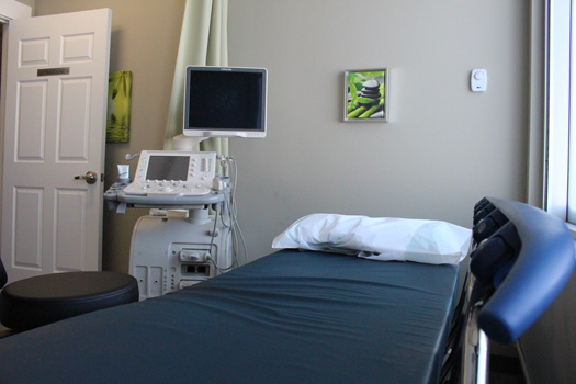
What Happens During My Echocardiogram?
- We will weigh you so we can index your heart size to body height and weight.
- Your sonographer may ask you to change into a gown.
- You will be taken into the examination room and will have three leads placed on your chest. Placement will be done as discreetly as possible for your comfort.
- Next, you will be instructed to lie down, and the sonographer will apply a warm, hypoallergenic ultrasound gel to your chest.
- The sonographer will begin scanning your heart with the transducer. At some point, the sonographer will have to apply some pressure to the middle and left side of your chest and under your breast. Some people find this part of the exam to be a little uncomfortable.
- You can expect to hear lots of noises from the machine during this exam. This is the Doppler technology assessing blood flow through your heart.
- The sonographer may also ask you to breathe in a certain way during the exam.
- After the sonographer has captured enough images, you are free to leave.
- A radiologist will review the images and send a detailed report to your doctor, usually within one business day.
Orientation
During an echocardiogram, your sonographer will position you in two different ways on a special bed.
Initially, you will be on your side, with one arm extended over your head. The sonographer will press the transducer against your upper rib cage through a cut-out on the special bed. They will also press the transducer against your chest and solar plexus.
The second position is on your back. The sonographer will press the transducer against your upper abdomen, lungs, and the notch in your neck. This position gives our radiologist an alternative viewing angle of your heart and its anatomy.
Cost
If you have an Alberta Health Care card or valid health care card from out of province, there is no cost for an echocardiogram (except in Quebec).
Duration
An echocardiogram lasts approximately 45 to 60 minutes.
Exam Preparation
Being prepared for your heart ultrasound helps us take the best possible images for diagnosis. Visit our exam prep page for more instructions specific to echocardiography.