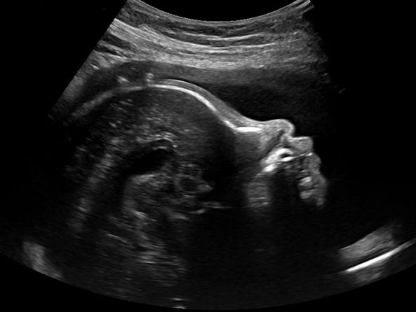
Obstetric Ultrasound
Obstetric ultrasound, also known as prenatal or pregnancy ultrasound, uses high-frequency sound waves to produce images of a developing embryo or fetus. The procedure also monitors the health of the mother’s uterus, ovaries, and the blood flow through the umbilical cord to the placenta. Your doctor will use information from obstetric ultrasounds to track pregnancy progress, gestational age and help predict delivery dates.
Reasons for an Obstetric Ultrasound
Your doctor may refer you for an obstetric ultrasound to:
- Confirm fertilization, implantation, and the presence of an embryo
- Identify whether it is a single or multiple pregnancy
- Estimate the fetus’s age and the due date
- Evaluate the position of the fetus and placenta in relation to the birth canal
- Check the amount of amniotic fluid around the fetus
- Monitor fetal development
- Check for congenital disabilities, hereditary conditions, or abnormalities
Types of Obstetric Ultrasound Scans:
What Happens During my Obstetric Ultrasound?
- The sonographer will apply a warm, hypoallergenic ultrasound gel to your abdomen and move a transducer around your belly to gather images of the fetus.
- The sonographer will move the transducer around the target area with moderate pressure to obtain images. This pressure should not cause pain. Please inform your sonographer of any discomfort.
- If you are having trouble holding your bladder or are in an uncomfortable position, talk to your sonographer so we can make adjustments.
- During your scan, the sonographer will be concentrating on completing your medical exam in a timely and coherent manner; therefore, try to save your questions for the end of the exam.
- At the end of your exam, we will print some ultrasound images, and we can also send digital photos to your cell phone or email for maximum convenience.
- A radiologist will review the images and send a detailed report to your doctor, usually within one business day.
Important Notes
- Sometimes you will be scheduled for additional scans. These follow-up scans do not mean there is a complication with your baby! Sometimes the gestational age or position of the fetus inside the uterus makes it challenging to take the right photos for diagnosis. Usually, in these situations, we will schedule a follow-up appointment.
- Pictures or videos are not allowed inside the exam room. Please respect the privacy of our staff and the sensitive nature of the health care industry.
- Our sonographers will gladly provide you with multiple baby photos over text or email and/or by printing the images.
Orientation
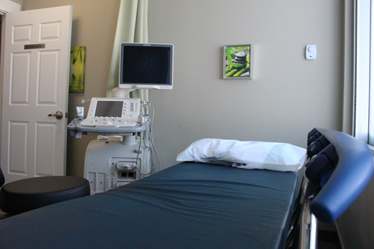
During an obstetric ultrasound, you will be on a bed, usually on your back. You may be asked to change position during your scan. Changing position helps move your organs and the fetus into a better position so the sonographer can capture high-quality images.
Cost
If you have an Alberta Health Care card or valid health care card from out of province, there is no cost for an obstetric ultrasound (except in Quebec).
Duration
An obstetric ultrasound scan lasts approximately 45 to 60 minutes. If you have multiple fetuses, the exam will take longer, usually about 90 minutes.
Support from Significant Others
One significant other is welcome to join you in the room for the entire duration of the exam. Your plus-one can be anyone important in your life who you want to share the experience with, such as your husband, wife, girlfriend, boyfriend, mother, sister, etc.
If you have more family who would like to attend the scan, our sonographers are willing to bring them into the room near the end of the exam to show them images of the baby. However, we do not allow more than one person in the room for the entire duration of the exam.
This policy allows us to support our employees by creating a comfortable work environment where they can perform their duties to the best of their abilities. As a patient, you must remember that any obstetrical evaluation is still a medical exam.
Occasionally, expecting mothers will bring their young children to the appointment, as they are not able to find a sitter. While we allow children in the exam room, we ask that you bring toys or activities to keep them occupied. We supply colouring books and crayons, but your child might prefer a game or movie on a tablet or iPad. Our goal is to create a stress-free environment for our employees as well as our expecting mothers.
Please Save Your Questions Until the End of the Scan
Pregnancy is both an exciting and stressful journey, and you have lots of questions. We ask that you save your questions for the sonographer until the end of the exam. Your sonographer will answer any questions within their scope of responsibility, but the majority will likely need to be answered later by your referring doctor.
While this may seem insensitive, please understand there are many reasons this policy is in place:
- Your sonographer’s job is to capture images of the highest quality, not to interpret them.
- Legally, health care technologists are not permitted to discuss results with the patient.
- Resulting images are reviewed by our radiologists, who have over 13 years of post secondary education plus additional years of specialization and industry experience.
- Our radiologists forward their reports to your doctor, who will often combine their information with results from additional tests and your patient history to form a comprehensive diagnosis.
- Your doctor will review everything with you in private and determine the next steps in your health care journey.
Please help us respect the relationship you have with your doctor, and allow our staff members to do their jobs to the best of their ability.
Ultrasound Baby Pictures
After your exam is complete, our sonographer will ask if you would like any images (sonograms) from the exam.
At Insight Medical Imaging, we can provide photos in three ways:
- Physical copies printed at our clinics
- Digital copies texted to your mobile device
- Digital copies sent to your email
We encourage patients to take both digital and physical copies of their ultrasound photos. Digital photos are easier to share with friends and family, and you don’t have to worry about losing or damaging them.
Twins or Multiples
Women with a multiple pregnancy require more frequent checkups and ultrasounds to monitor health, growth, and fetal development. The only way to confirm a multiple pregnancy is with ultrasound imaging.
Detection of Multiples
Multiples may not be detected in the first trimester. There are rare situations in which the second embryo is “hidden” during a dating ultrasound around the six- to seven-week mark. Your sonographer may not detect a second embryo due to fetal positioning or a lack of development.
After the 20-week mark, during the second trimester, a second fetus will typically be easily identifiable on an ultrasound.
What About a Second Heartbeat?
One of the first steps in most ultrasounds is checking your baby’s heartbeat. Naturally, it makes sense to check for a second heartbeat to confirm a second embryo. Unfortunately, many variables impact the detection of a second heartbeat. For example:
- Your own heartbeat can complicate the detection process.
- Varying development timelines mean some heartbeats are hard to detect until the end of the first trimester.
Impact of Multiples on Scan Time
It is important to understand the impact multiple fetuses have on ultrasound scan times. Insight Medical Imaging books 45-minute time slots for each obstetrical ultrasound. If you are having twins, that time frame doubles to a 90-minute time slot. If you have triplets, we will set aside 135 minutes for your exam.
However, just because we book you in for 45, 90, or 135 minutes does not mean your exam will take that long. Exam time varies significantly from patient to patient.
Obstetric Ultrasound Side Effects and Risks
To date, the consensus is that diagnostic ultrasound has shown no harmful side effects to pregnant mothers or their fetuses (Joy et al., 2006).
The procedure is non-invasive and immediate and does not involve exposure to ionizing radiation. While studies have been conducted for decades on the biological, mechanical, and thermal effects of high-frequency sound waves used in ultrasound imaging, no clinically significant findings impacting human health have been demonstrated (Abramowicz, 2013).
Nevertheless, Insight Medical Imaging practices “as low as reasonably achievable" (ALARA) principles with ultrasound imaging, just as we do with all our services. Therefore, we do not recommend ultrasound imaging unless there is a medical goal or diagnostic purpose. As a precaution, we ensure our sonographers always use the minimum acoustic power needed to generate optimum images for diagnosis.
Obstetric Ultrasound Scans:
Dating Ultrasound
Dating ultrasound scans, also known as early ultrasounds, are typically the first type of obstetric evaluation during pregnancy. These ultrasound scans are usually performed between 7 and 12 weeks of pregnancy.
Purpose of a Dating Ultrasound
The goal of a dating ultrasound is to establish the gestational age of the pregnancy. Our sonographer will calculate gestational age by measuring the fetus from crown (head) to rump (buttocks).
Once your doctor establishes your pregnancy timeline, they can provide more accurate prenatal care instructions. These scans also provide expecting couples with an approximate due date that is, as the Society of Obstetricians and Gynaecologists of Canada states, usually accurate to within five days.
Is a Dating Ultrasound Internal or External?
Sonographers can perform dating ultrasounds with an external (transabdominal) or internal (transvaginal) transducer. There are numerous factors, such as body build, that impact transducer selection; however, the most important factor is the size of the embryo.
Physicians typically order a dating ultrasound around the 7- to 12-week mark. If your appointment is around the 7-week mark, there is a chance the developing embryo might be too small to see with an external transabdominal probe. In this situation, our sonographers ask for permission to perform a transvaginal (internal) scan.
If permission is granted, the sonographer will hand you an ultrasound wand that you may gently insert into your vagina. We do our best to make this experience as comfortable as possible.
While the majority of our sonographers are female, we are an equal-opportunity employer that hires sonographers of both genders. Should you require a transvaginal examination and your sonographer is male, we ensure a female colleague is in the room with you for the duration of the internal scan.
Dating Ultrasound Accuracy and Expected Date of Delivery (EDD)
Dating ultrasounds provide precise crown-rump length (CRL) measurements to determine gestational age and EDD. Ultrasound scans performed within the 7- to 12-week period provide the highest due date accuracy, as the fetus is in the best position for measuring.
The fetus is in the most linear position during this time frame, growing rapidly but unable to twist or alter its body position significantly. Therefore, an ultrasound during this period provides the most accurate crown-to-rump measurement for predicting delivery.
EDD Using Last Menstrual Period (LMP) vs. Dating Ultrasound
Your doctor will determine your EDD using either your LMP or your dating ultrasound. But how do they choose between the two methods?
- Typically, your doctor will use your LMP if it has been reliably identified. For it to be considered reliable, the EDD from your dating ultrasound must be within five days of the EDD from your LMP.
- Sometimes your doctor will use your dating ultrasound to determine your due date. This method is chosen when your LMP is unknown or the EDD from your dating scan differs by more than five days from the EDD calculated with your LMP.
- Your doctor might also rely on your dating scan if your cycle is irregular or if you have recently been on a birth control pill. Both factors can affect ovulation and conception timelines, making your LMP less reliable.
Sometimes people are given conflicting delivery dates. While you might think the logical solution is to rely on the most recent estimate, the opposite is true. Ultrasound examinations during the 7- to 12-week time frame are the most accurate, to within five days.
Scans from the 12- to 22-week period are accurate within 10 days, and anything past the 22-week mark is considered unreliable. After 22 weeks, the gestational age of a fetus is too hard to predict because it starts showing individual growth potential. Differences in development and size can skew gestational age estimates by as much as two or three weeks.
First-Trimester Combined Screening (FTS)
FTS is a test for chromosome conditions and congenital disabilities. It is available to all women between 11 and 13 weeks plus six days of pregnancy. With FTS, two measurements are used, together with your age, to estimate your chance of having a baby with Down syndrome, trisomy 18, or trisomy 13. The first measurement is the nuchal translucency (NT) measurement, taken by ultrasound, and the second is a blood test.
Nuchal Translucency (NT) Measurement
NT ultrasound exams are performed between 11 weeks and 13 weeks plus six days of pregnancy. During an NT exam, your sonographer will measure the fluid at the back of the Fetuses neck (called nuchal translucency). All Fetuses have some fluid here. Fetuses with Down syndrome are more likely to have an increased amount of fluid.
The NT ultrasound also:
- Confirms that the baby has a consistent heartbeat
- Confirms your dates
- Diagnoses a multiple pregnancy
- Checks for other congenital disabilities
The Maternal Blood Test
The maternal blood test measures two substances, PAPP-A and free beta-hCG, that are usually found in the blood of all pregnant women. When Down syndrome is present, the levels of free beta-hCG and PAPP-A tend to be out of the expected normal range.
How Accurate is an FTS Exam?
FTS has an 85 to 90 percent detection rate. Therefore, the FTS exam will identify an increased risk of Down syndrome, trisomy 18, or trisomy 13 in 85 to 90 percent of actual cases (Loncar et al., 2010).
What Type of Results Should I Expect from FTS?
Your doctor will provide a personal risk estimate that tells you how likely it is that your pregnancy will be affected by Down syndrome, trisomy 13, or trisomy 18. In the FTS report, this estimate is called the “adjusted risk.”
What Happens After FTS?
Your FTS results help your doctor decide on the next course of action. Together, you may opt for additional prenatal testing, such as amniocentesis or chorionic villus sampling (CVS).
Most women receive reassuring results and do not need further testing, other than the routine 18-week anatomy scan offered to everyone.
Women who receive a risk result indicating that their fetus is at an increased risk for Down syndrome, trisomy 18, or trisomy 13 are offered the option of having a diagnostic test, such as amniocentesis.
Regardless of a woman’s age or FTS results, the decision to have a diagnostic test is a personal one.
Amniocentesis and Chorionic Villus Sampling (CVS)
Samples collected from these tests contain the baby’s cells. Laboratory technicians look at the chromosomes within these cells to determine whether the baby has a chromosome condition.
These procedures are not routinely offered to all women, as they increase the risk of miscarriage. Most women undergoing amniocentesis or CVS do not have complications following the procedure and receive reassuring chromosome results.
Benefits of FTS
- Early, more accurate screening gives peace of mind to many women.
- The FTS personal risk estimate can be used to help women make more informed choices about diagnostic testing.
- It is possible to detect specific, significant congenital disabilities at the time of the NT scan.
Limitations of FTS
- About 1 in 20 women will receive an increased-risk result. It is normal to be worried when you hear your fetus is at an increased risk. Most women with increased-risk results do have healthy babies.
- An increased-risk result does not mean that the baby has a chromosome condition.
- A reduced-risk result does not guarantee a healthy baby
Anatomy Ultrasound
Anatomy ultrasounds happen during the second trimester, usually around the 18- to 22-week mark. Sometimes these scans are referred to as a level-two ultrasound or the 20-week anatomy scan. The anatomy scan is usually the most memorable obstetric ultrasound for parents because it is the first time they get to see their baby.
Purpose of an Anatomy Ultrasound
The goals of an anatomy scan are to examine the fetus’s anatomy and heart rate, measure its growth, evaluate the placental position in relation to the cervix, and monitor amniotic fluid volumes. When examining anatomy, the sonographer will look at the brain, face, abdomen (kidneys, stomach, bladder, liver), lungs, diaphragm, spine, sexual organs, and all four heart chambers.
Determining Gender at the Anatomy Ultrasound
If your sonographer can identify sex organs at your anatomy scan, it is highly likely that they can tell you the gender of your baby. Please inform the sonographer at the beginning of the exam if you do or do not want to learn the potential gender of your baby.
Also, note that the fetus’s sex organs are not the primary area of interest during an anatomy scan. During this scan, the sonographer is looking for critical medical information such as heart rate, brain development, spinal structure, etc.
Sometimes fetal genitalia cannot be seen during an anatomy scan due to body position. If the fetus has its legs crossed or is facing away from the abdomen, it can be challenging to determine the gender.
Depending on how long the medical portion of your scan takes, our sonographers may be able to work with you to get the fetus into a better position to estimate gender.
If there is enough time left after gathering the medical images needed by the radiologist, we may give patients a juice box or snack and ask them to walk around the clinic. The extra sugar and liquid from the juice box combined with walking may encourage the fetus to change position, which can help our team identify sex organs.
Gender Identification Is Never 100 Percent Accurate
Should you want to learn the gender of your fetus, you must remember that identification is never 100 percent accurate. Many factors can influence gender identification, including:
- Fetus position
- Movement restricted by the size of the uterus
- Testicles that have not descended, making the fetus look female
- Ambiguous genitalia (baby may have a mix of both reproductive organs)
- Clarity of the images and skill of the sonographer
Biophysical Profile (BPP) Ultrasound
A BPP ultrasound is commonly performed any time after 28 weeks of pregnancy, in the third trimester.
Purpose of a BPP Ultrasound
The goal of a BPP ultrasound is to assess fetal well-being leading up to the estimated delivery date. This precautionary exam monitors the supply of oxygen to your baby. If oxygen is ever compromised, your doctor may induce labour or consider a Caesarean section to protect your baby.
Who Typically Needs a BPP Ultrasound?
Your healthcare provider may recommend you for a BPP if you:
- Do not deliver on your due date
- Are past 40 weeks’ gestation
- Are carrying twins or multiples
- Have high blood pressure or type 2 diabetes
- Have a history of pregnancy complications
- Are older than 35
BPP Ultrasound Frequency
Since BPP scans are performed to monitor the health of your baby, your doctor may order multiple scans in your third trimester. It is not uncommon to be sent on a bi-weekly basis as you near the 40-week date. However, you may not be sent for any BPP scans during your pregnancy. You will work with your doctor to determine what is best for you and your baby.
What Attributes Does a BPP Ultrasound Assess?
Typically, a BPP scan assesses five attributes:
- Fetal heart rate
- Fetal breathing
- Fetal body movements
- Fetal muscle tone
- Amniotic fluid volume
What Happens After the BPP Procedure?
Our team will record your test results and send them to your doctor. Your doctor will assess your baby’s health based on heart rate, breathing, body movements, muscle tone, and amniotic fluid.
Depending on the score, your doctor may not take any additional action. You will continue with your pregnancy, waiting for labour to start naturally.
Your doctor may book additional scans for precautionary reasons. Alternatively, they may schedule induced labour or a Caesarean section if they feel your baby’s health may start to be affected.
Complete Series
When talking to your doctor about your first ultrasound appointment, be sure to ask about Insight’s “complete series” imaging option. We created this option after receiving feedback from referring physicians.
If your doctor selects the complete-series option, our team will book you for the three primary ultrasound appointments (dating, nuchal translucency, and anatomy) simultaneously so you don’t have to stress about rebooking at the appropriate time.
Should you require additional routine imaging, a biophysical profile scan, or any other scans, we are happy to oblige. The complete-series option makes life easier for you and your doctor, as you have fewer appointments to book.
Exam Preparation
Being prepared for your obstetric ultrasound helps us take the best possible images for diagnosis. Please visit our exam prep page for more instructions specific to obstetric ultrasound.